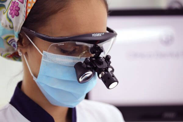
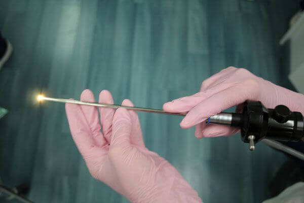
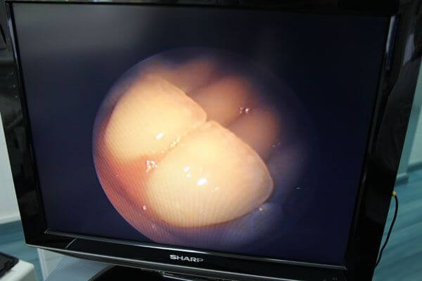
Surgical microscopes, such as the self-illuminated microcameras on endoscope equipment, provide us with very useful, detailed views of the hidden facets of dental anatomy.
We can, for example, see the inside of the tooth and the root canals at large magnifications. This is of great help when determining the diagnosis and treatment of complex situations. It assists us when working in different areas of dentistry, such as conservative dentistry (detection of fissures, hidden caries, etc.), endodontic treatment (locating root canals, elimination of pulp calcifications, re-treatments, etc.) and surgery (microsurgery, implants, grafting, complicated extractions, etc.), amongst others.


When we are able to see the details at greater magnifications, we can provide more accurate diagnoses and select treatments with more precision.
One of the biggest advantages of these systems is the fact that they use coaxial light. This means that the light shines in exactly the same direction as the camera or the lens which is observing it and, therefore, such systems do not generate shadows, unlike in simple observations where the light shines at a different angle from the line of sight.
This translates into finer and more accurate diagnoses and treatments.

Our different optical magnification systems, such as microscopes, surgical loupes or endoscopes, result in more accurate diagnoses and treatments


