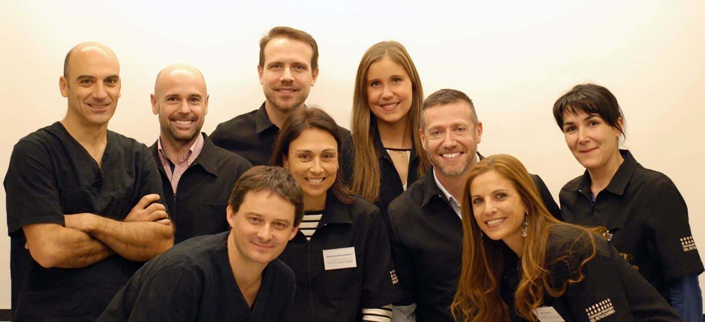
From November 20th to November 21th we’ve travelled to Madrid to attend the microscopic dentistry course given by Dr. Christian del Rey and Dr. Guillermo Carrera. We’ve shared the experience with an excellent group of professionals and we’ve learned a lot of practical tips on the use of the dental microscope. The use of the dental microscope allows us to use microtreatment and microsurgery techniques with a high level of detail. It also allows us to work in a very ergonomic position, which avoids fatigue to the doctors and prevents lesions due to working posture.
The dental microscope is quite difficult to use. It’s much more difficult than in other medical specialities, like ophthalmology or neurosurgery, as an example, due to the fact that the access to different surfaces inside the mouth can be very complex. The three main difficulties come from the limited visual depth of field given by the high magnification objectives, the reduced visual field, which forces the patient to stay without moving and the access to small cavities inside the mouth which makes us work very often through an specular vision (using dental mirrors). The view through the mirrors while using the dental microscope flips the image that we see, so that when we see something up, in fact it’s down and when we see it down, in fact it’s up. So a lot of training is needed by the dental clinician to have absolute control of the more precise techniques.
On the other hand, using augmented vision on dentistry allows us to work without damaging our back or neck and allows us to see very fine details not only due to the magnification, but also due to the coaxial illumination (a light source that goes in the same direction of the vision line. This helps us to see, for example, the small space under the gums or the inner part of the dental nerve canals with great definition.
The course has been very practical and we really had very nice moments with all the group.
